
SIMDALEE2 – MidTerm Status
Over the past century, electron microscopy (EM) has played a paramount role in making the invisible visible. The tremendous impact of this technique on the life sciences, knowledge about processes in the environment on earth as well as other bodies in the universe are nowadays widely appreciated by the general public. On the other hand, for individuals involved in technological developments in industry as well as in fundamental research, EM-techniques are indispensable tools, accessible either in specialised research laboratories or large scale research facilities. Conventional EM-techniques derive their resolving power in the sub-Ångstrøm (!) range from high acceleration voltages of typically a few hundred thousand volts. In recent decennia, electron optics for low energy electrons (LEE, several volts up to several hundred volts) have been developed with a resolution in the nm-range, which is still acceptable for a broad variety of applications. This not only allows construction of table-top design electron microscopes, but also offers the additional advantage of less destructive investigations, particularly important for soft-matter investigations and life science.
Essential steps in the earlier development of LEE technology were taken by members of the SIMDALEE2 consortium, which – after recruitment of a group of 14 young researchers – has established an intersectoral and interdisciplinary network striving to develop compact and affordable electron microscopy systems based on low energy electrons which can be put to more widespread use.
How does the support by the European Union of such a large scale venture merit society? In this connection one might think, for example, of targeted therapy e.g. for women suffering from HER-2 positive breast cancer. Presently, the patient’s response to treatment is often only monitored à posteriori through the effect the drugs take on her general condition. Here the availability of a compact and perhaps dedicated EM-system monitoring the drug-response on a microscopic level, could help, not only in clinical research, but, even more importantly, in therapeutic deployment to optimally tailor the therapy to the individual patient’s needs during treatment.
To achieve the goal mentioned above, the SIMDALEE2 consortium is not only taking steps in technological development of LEE-techniques, but is also addressing the needs to further fundamental understanding of the underlying physical processes involved. The latter concerns the following processes: (1) field emission from a tip used as high-brightness LEE-source in a microscope; (2) the interaction of LEEs with solids; (3) the mechanism leading to creation of secondary electrons (SEs); and (4) the subsequent emission of SEs from a solid surface. The interaction of LEE with solids is not only important for quantitative understanding and interpretation of the signal in electron microscopy. In biological tissue, the passage of LEE also plays an important role in damaging DNA leading to the development of tumors, while LEEs are at the same time presently being considered as therapeutic agents for tumor treatment. Finally, the transport of LEE in matter is also of paramount technological interest, such as in present and future semiconductor technologies where creation and penetration of LEEs in photoresists often limits the achievable level of miniaturisation. Furthermore, a better understanding (and perhaps even control!) of the generation and emission of SEs is needed for particle detectors, accelerator physics, in particular the next generation high luminosity large hadron collider (HL-LHC), space technology and nanotechnology in general.
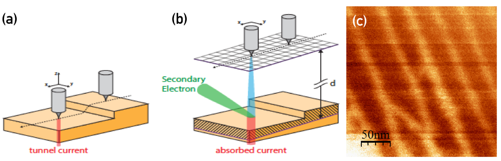 Schematic illustration of the principle of the near-field emission secondary electron microscope (NFESEM). (a) common Scanning Tunnelling Microscope (STM); (b) In the NFSEM-mode the tip is retracted from the surface by about 1-10 nm. Application of a bias voltage leads to field emission from the tip. The secondary electrons created in the sample or the absorbed current as a function of the position of the tip lead to the recorded NFSEM image; (c) NFESEM image of a stepped W(110) surface.
Presently, the efforts in developing prototypical LEE-technology focus on the so called near field emission scanning electron microscope (NFESEM, see the figure above). A 3-dimensional theory beyond the Fowler-Nordheim approach is under development and put to a stringent test by comparing NFESEM-measurements with theoretical calculations using the electric field around a specific tip measured with electron holography. In order for a meaningful comparison to be possible, the very tip used in the NFESEM measurements is transported in a specially constructed ultra-high-vacuum suitcase to the electron microscope where the holography measurements are carried out. The generation and emission of SEs in the target and the passage through the sample-tip electric field to the electron detector are also being theoretically modelled. The latter model is individually tested by comparison with so-called secondary electron–electron energy loss coincidence spectra (SE2ELCS) which provides detailed information about the SE-generation and emission mechanism. The parameters dominating SE-emission over the surface barrier are also addressed by various experimental approaches across the network. In this way, the entire signal generation process in the NFESEM according to the present theoretical understanding can be compared to experiment on a microscopic level. Furthermore, a suitable electron detector as well as field emission tips based on novel materials are being developed. Additional experiments are being set up to enhance quantitative understanding concerning the interaction of LEE with solids.
While the above activities are presently underway within the projected timescale, a highlight that should be mentioned here in conclusion is the successful measurement of the magnetical hysteresis-loop of a sample consisting of a ferromagnetic model system (a few monolayers of Fe on a W (110) specimen), which was magnetised by a set of small coils in Helmholtz-configuration inside the NFESEM experimental chamber. For this purpose, the signal electrons in the NFESEM were subjected to spin-analysis using a Mott-detector. This demonstrates that not only morphological and chemical information on the nanometer-scale can be obtained with LEE-technology, but that magnetic imaging can also be performed with the same spatial resolution and on the very same spot of a specimen!
Aditional information can be obtained by contacting the project coordinator, Prof. Wolfgang Werner (werner[at]iap.tuwien.ac.at).